Gallium Tumor Imaging:
Gallium tumor localization is likely multifactoral, but in part related to leaky capillary membranes in tumors, and the presence of iron-binding proteins such as ferritin which are found in increased concentrations in tumors.
Bronchogenic carcinoma:
Squamous cell carcinoma has the highest detection rate (gallium avidity), while adenocarcinoma has the lowest. Overall, gallium has a sensitivity of about 90% for the detection of primary bronchogenic carcinoma. Scatter from adjacent structures reduces the detection rate of lesions near the mediastinum and near the liver. Lesions smaller than 1.5 cm are also difficult to detect. Lack of gallium accumulation in a lesion is still associated with a 24% probability for malignancy. When a primary tumor accumulates gallium, the likelihood that an extrapulmonary site of uptake represents a metastatic lesion is about 90%.
Lack of mediastinal uptake in a patient with a gallium-avid primary tumor is associated with a less than 50% likelihood of mediastinal disease, but the use of gallium for this purpose is not widespread and remains controversial.
Hepatocellular Carcinoma:
Approximately 90% of hepatomas are gallium avid. About 50% demonstrate activity greater than the adjacent liver, 30% have uptake equal to the surrounding liver, and 10% have uptake which is less than the adjacent liver. For these reasons, it is essential to view the gallium exam in conjuction with a Tc-Sulfur colloid exam. Cold lesions on the sulfur colloid study which "fill-in" on the gallium scan are highly suspicious for hepatoma in the appropriate clinical setting. Overall sensitivity is reported between 85-90%.
Lymphoma:
1. Hodgkin's:
Gallium scanning can be used in patients with Hodgkin's lymphoma in order to determine tumor avidity for Ga-67, determine the extent of disease, for the evaluation of a residual mass, to monitor response to treatment, and to predict disease free and over-all survival. About 90% of Hodgkin's lymphomas (Nodular sclerosing, Mixed cellularity, and Lymphocyte depleted which account for about 90% of tumors) are gallium avid pre-treatment. Lymphocyte predominant tumors (10% of cases) show somewhat less gallium avidity (79%). Overall sensitivity for detecting Hodgkins lymphoma is about 85%, with a specificity of 90%. SPECT has a sensitivity of 95% and a specificity of 90% for the detection of mediastinal disease in Hodgkin's lymphoma. Mediastinal or hilar nodal involvement is more common at presentation in patients with Hodgkin's lymphoma (75%) than in non-Hodgkin's lymphoma (25%). Nodal involvement of the hila and mediastinum is usually asymmetric. The gallium exam is not useful for assessing splenic involvement due to normal uptake in the spleen. The periportal area may also be difficult to assess. Bone involvement is usually well demonstrated and persistent uptake following treatment indicates the presence of active disease [13].
Gallium scintigraphy can also be used to predict patient response to treatment after initiation of chemotherapy [7]. Normalization of a positive pre-therapy scan (ie: a negative scan) after one cycle or at mid cycle is associated with a high likelihood of complete response (82-92% of patients) [7]. A persistently positive scan after one cycle of chemotherapy may indicate a need for a different treatment protocol [7]. Because chemotherapy may suppress gallium uptake by the tumor, scans should be performed at least 2 weeks after the cycle of chemotherapy [7].
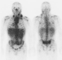
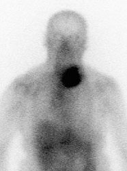
Hodgkins Lymphoma: The Ga67 scan below demonstrates nodal uptake of tracer in the neck and mediastinum in this patient with Hodgkins disease. Click image to enlarge. |
|
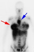
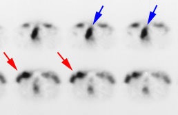
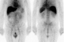
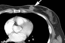
Hodgkins Lymphoma and Mastitis: The patient shown in the images below was a post-partum lactating female who presented for a baseline Ga-67 exam as part of her staging work-up for Hodgkins lymphoma (nodular sclerosing subtype). The planar whole body images demonstrate abnormal uptake of gallium within the patient's primary tumor in the mediastinum (blue arrow). Breast uptake is seen bilaterally (consistent with the patients history of lactation), but is asymmetric- right greater than left (red arrow). SPECT imaging was also performed (right). Asymmetric breast uptake was concerning for lymphomatous involvement of the breast, but on examination the patient was found to have a right breast mastitis. (Click images to enlarge) |
|
|
Ga-67 in Recurrent/Residual Disease:
Gallium is very useful in the detection of recurrent or residual disease. When scanning patients for recurrence it is essential to image the entire body since about 25-27% of patients will relapse only in new disease sites different from the primary site of involement [1,2]. Up to two-thirds of patients with initial mediastinal involvement will have persistent radiographic mediastinal abnormalities following completion of therapy [10]- yet only 18% of these patients ever relapse [2].
When scanning these patients it is important to remember that chemotherapy may suppress gallium uptake for some time after treatment in spite of active disease [2]. At least a 3 week delay after completion of chemotherapy is recommended prior to re-imaging [2]. Tumor irradiation may also result in transient or permanent loss of gallium uptake. Since gallium is a viability agent, it is taken up by cancer tissue and not by fibrotic or necrotic tissue at the tumor site [1]. When gallium uptake is observed in a residual mass indicating viable tumor following treatment, further non-cross resistant treatment should be instituted as gallium imaging has an overall accuracy of up to 93% for active disease [10]. The presence of residual gallium uptake after treatment has been shown to be a poor prognostic indicator in patients with Hodgkin's and non-Hodgkin's lymphoma [2]. In Hodgkin's patients overall survival decreased from 100% to 60% at 2 years in patients with positive scans at the end of treatment and decreased from 90 to 35% in non-Hodgkin's patients [2].
Hilar nodal activity can be seen in 50-85% of patients that have completed chemotherapy and can persist for up to 4 years on follow-up exams (mean 27 months) [9]. The finding may be evident on SPECT images only in about one third of these cases. Bilateral symmetric hilar activity that is less intense than the original disease is essentially benign and is only very rarely associated with the presence of residual lymphoma. Hilar activity may be related to the presence of a concomitant inflammatory disease or possibly a direct effect of the chemotherapy- although the persence of hilar activity has no correlation with dosage received [9]. Asymmetric or unilateral hilar uptake post therapy is somewhat more concerning and should raise the possibility of recurrent disease- especially if the intensity is similar to the original disease [9]. If necessary, thallium imaging may be useful in these instances as it will accumulate within tumor involved nodes, but will be negative in the absence of disease.
Diffuse lung uptake of Ga-67 has also been described following treatment, occurring in up to 12% of patients. The uptake is most commonly mild and does not indicate lymphomatous involvement of the lungs. The uptake most likely reflects pulmonary toxicity secondary to chemotherapy [3].
There may be utility to performing imaging during the patients initial treatment. A complete response is achieved in 70% of patients during the first 3 cycles of treatment. These patients also demonstrate longer disease free periods than patients who require more cycles to achieve a complete response.
2. Non-Hodgkins:
Gallium sensitivity is reported to be better than 85% for high grade tumors, and is particularly good for Histiocytic lymphoma (89%). Patients with high grade tumors generally have a poor prognosis. Sensitivity for low grade (slow growing) tumors is poor, although these patients tend to have a favorable prognosis. Detection of well differentiated lymphocytic lymphoma is also poor (60%).
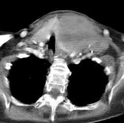
Non-Hodgkins Lymphoma: The Ga67 scan below was performed to evaluate for other sites of disease in a patient with a large non-Hodgkin's lymphoma in the left neck. Intense tracer uptake can be seen in the patient's tumor, but there were no other sites of disease identified. |
A persistently positive Ga-67 exam after one cycle of treatment or at midtreatment for non-Hodgkins lymphoma is associated with a higher likelihood for treatment failure [8], while a negative scan implies a favorable prognosis [6]. In patients with positive Ga67 scans after one cycle and at midtreatment, 71% and 74%, respectively, had treatment failure [8]. Negative scans after one cycle and at midtreatment were associated with complete response in 81% and 63% of patients, respectrively [8]. A persistently positive scan during treatment may be cause to justify a change in therapy [6,8]. The earlier an ineffective treatment is stopped and a more appropriate one instituted, the better the chances for tumor response and longer failure-free survival [8].
Thallium imaging can add useful information in the evaluation of Non-Hodgkin's lymphoma. Thallium generally has a higher avidity for low grade lymphomas than gallium, while its uptake in high grade lesions is usually poor (approximately 25% of low grade lymphomas are thallium positive and gallium negative). Thallium scanning in the evaluation of non-Hodgkin's lymphoma is best performed at 1 hour post-inject in order to improve target to background activity. Due to gut activity, thallium imaging is not useful for the evaluation of abdominal or pelvic disease.
For both non-Hodgkin's and Hodgkin's lymphoma the response of sites of osseous involvement to treatment are best monitored with Ga-67, as the bone scan may demonstrate persistent scintigraphic abnormalities, even following successful therapy [4].
3. Burkitts:
Of Burkitts lymphomas, 90% or greater are gallium avid.
Melanoma:
About 75% of lesions over 2cm in size are gallium avid. For lesions less than 2 cm in size, only about 20% show gallium uptake. Overall sensitivity and specificity have been reported to be 82-90% and 99% respectively.
Mesothelioma:
Gallium has been found to be more sensitive than chest x-ray in differentiating
malignant mesothelioma from benign pleural thickening, but experience is limited.
Soft tissue sarcoma:
Gallium has a high sensitivity (about 85-90%) for detecting active sites of disease. Gallium uptake also tends to correspond to the tumor grade. A gallium avid site that becomes negative after therapy is indicative of a favorable clinical response.
Testicular Carcinoma:
Gallium uptake in metastatic testicular carcinomas varies according to the cell type with a sensitivity of about 90% for seminomas, 75% for embryonal cell, and only 25% for teratomas. Gallium scanning can be used to distinguish post therapy fibrosis from residual active disease in patients with mediastinal and para-aortic metastases [5]
REFERENCES:
(1) J Nucl Med 1993; Front D, et al. Early detection of lymphoma recurrence with
gallium-67 scintigraphy. 34: 2101-04
(2) Seminars in Nucl Med 1995; Front D, Israel O. The role of Ga-67 scinitgraphy in evaluating the results of therapy of lymphoma patients. 25: 60-71
(3) Radiology 1996; 199: 473-476
(4) J Nucl Med 1995; Bar-Shalom R, et al. Gallium-67 scintigraphy in lymphoma with bone involvement. 36: 446-50
(5) J Nucl Med 1994; Uchiyama M, et al. Gallium-67-citrate imaging in extragonadal and gonadal seminomas: Relationship to radiologic findings. 35: 1624-30
(6) J Nucl Med 1998; Gaasparini M, et al. Gallium-67 scintigraphy evaluation of therpay in non-Hodgkin's lymphoma. 39: 1586-1590
(7) Radiology 1999; Front D, et al. Hodgkin disease: Prediction of outcome with Ga-67 scintigraphy after one cycle of chemotherapy. 210: 487-491
(8) Radiology 2000; Front D, et al. Aggressive non-Hodgkin lymphoma: Early prediction of outcome with Ga67 scintigraphy. 214: 253-257
(9) J Nucl Med 2000; Frohlich DEC, et al. When is hilar uptake of 67-Ga-Citrate indicative of residual disease after CHOP chemotherapy. 41: 269-274
(10) J Nucl Med 1992; Kostakoglu L, et al. Validation of gallium-67-citrate single-photon emission computed tomography in biopsy-confirmed residual Hodgkin's disease in the mediastinum. 33: 345-350
(11) J Nucl Med 1986; Tsan MF, Scheffel U. Mechanism of gallium-67 accumulation in tumors. 27: 1215-1219
(12) J Nucl Med Biol 1983l Berry JP, et al. Localization of gallium in tumor cells. Electron microscopy, electron probe microanalysis and analytical ion microscopy. 10: 199-204
(13) J Nucl Med 2002; Israel O, et al. Bone lymphoma: 67Ga scintigraphy and CT for prediction of outcome after treatment. 43: 1295-1303

